
Patients... Please Start Here.
Our advanced technology, highly-trained staff of board certified radiologists, and comfortable, patient-focused imaging centers give you peace of mind-and the confidence that you're receiving the best care available.
Using our state-of-the-art equipment, Concord Imaging Center's highly skilled technologists and physicians are able to conduct a variety of tests and procedures.
Our team then provides your physician's office with the most accurate and timely reporting, allowing for a thorough diagnosis to be made.
Along with the confidence that comes from knowing that your imaging team is outfitted with the diagnostic tools to provide better care for you, our investment in technology means you'll benefit from quicker, more comfortable procedures.
Our advanced technology equipment includes:
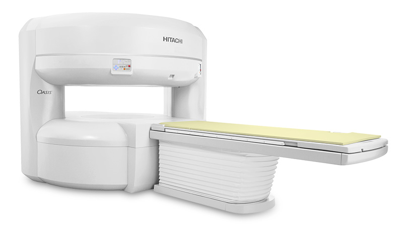
Philips Panorama High Field Open MRI
Our Panorama high field Open MRI appeals to both patients and physicians because it has a wide-open design, high image quality, large field of view and broad coverage of clinical applications. With these unique features, every patient, especially those who are anxious, elderly, or claustrophobic can have a relaxing MRI experience while at the same time benefitting from high resolution imaging.
 Sentinelle Vanguard Breast MRI Table
Demonstrating our continued commitment to leadership in the field of breast MRI , Concord Imaging Center is using the Sentinelle Vanguard high performance breast coil system with our GE High Field MRI in order to optimize image quality and position our patients for early breast cancer intervention. With features not available in traditional breast MRI coils, this next-generation system offers unprecedented improvements in image quality, ease of access for biopsies and patient comfort. Concord Imaging Center is one of a few places in the state where you can find this type of advanced technology.
Multidetector CT Scanners with dose modulation
Radiation exposure to the patient has become an increasing concern for patients and radiologists. Our state-of-the-art multidetector computed tomography (CT) scanners employ advanced automatic exposure controls (AEC) and dose modulation techniques to reduce the radiation dose to the patient while maintaining precision imaging.
Hologic Discovery™ Densitometry DXA Scanner
For women needing a Densitometry (DXA) scan, we offer the Discovery™ system, a single platform that supports a broad spectrum of patients over a lifetime of care. It allows us to assess not only vertebral fractures, but also provides visualization of abdominal aortic calcifications with a single scan. The Discovery™ scanner gives us the best diagnostic tools to support the early detection and treatment of osteoporosis and to monitor abdominal aortic calcifications, vertebral fractures and obesity.
Hologic Stereotactic-Guided Breast Biopsy Modality
When we tell our patients "this won't take long" at the beginning of their stereotactic-guided breast biopsy , we mean it. With the Hologic stereotactic-guided breast biopsy modality, total tissue acquisition time is less than 30 seconds with integrated pain management. This biopsy table is a vacuum-assisted breast biopsy system with the widest variety of needle sizes to address the broadest spectrum of patients and has a site marker, providing accurate delivery to the biopsy site.
Selenia Digital Mammography - R2™ ImageChecker® CAD
We are proud to offer digital mammography from Selenia with the R2™ ImageChecker® CAD (computer-aided detection), which helps our radiologists to better read the results of our patients' mammograms. After identifying regions of interest on mammography images, ImageChecker® CAD brings the regions to the attention of our radiologist, which in turn helps to decrease false negative readings. ImageChecker® CAD was the first FDA-approved mammography computer-aided detection system and has remained a leader in this type of technology by consistently delivering the best detection performance available.
McKesson Picture Archiving and Communication System (PACS)
A Picture Archiving and Communication System (PACS) is an elaborate network of computers that manage and deliver images and information. A PACS collects images from various types of radiology equipment (e.g. CT scan , MRI , ultrasound , etc.) and integrates them with health care organization information systems allowing for rapid transfer of patient data. A PACS also stores images in a secure environment, making them quickly and electronically accessible by authorized users when needed.